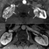
Renal Lesions in Autoimmune Pancreatitis: Diffusion Weighted Magnetic Resonance Imaging for Assessing Response to Corticosteroid Therapy
Abstract
Keywords
References
Chari ST, Smyrk TC, Levy MJ, Topazian MD, Takahashi N, Zhang L, et al. Diagnosis of autoimmune pancreatitis: the Mayo Clinic experience. Clin Gastroenterol Hepatol 2006; 4:1010-6. [PMID: 16843735]
Ghazale A, Chari ST, Smyrk TC, Levy MJ, Topazian MD, Takahashi N, et al. Value of serum IgG4 in the diagnosis of autoimmune pancreatitis and in distinguishing it from pancreatic cancer. Am J Gastroenterol 2007; 102:1646-53. [PMID: 17555461]
Sahani DV, Sainani NI, Deshpande V, Shaikh MS, Frinkelberg DL, Fernandez-del Castillo C. Autoimmune pancreatitis: disease evolution, staging, response assessment, and CT features that predict response to corticosteroid therapy. Radiology 2009; 250:118-29. [PMID: 19017924]
Kamisawa T, Egawa N, Nakajima H, Tsuruta K, Okamoto A. Extrapancreatic lesions in autoimmune pancreatitis. J Clin Gastroenterol 2005; 39:904-7. [PMID: 16208116]
Takahashi N, Kawashima A, Fletcher JG, Chari ST. Renal involvement in patients with autoimmune pancreatitis: CT and MR imaging findings. Radiology 2007; 242:791-801. [PMID: 17229877]
Sohn JH, Byun JH, Yoon SE, Choi EK, Park SH, Kim MH, Lee MG. Abdominal extrapancreatic lesions associated with autoimmune pancreatitis: radiological findings and changes after therapy. Eur J Radiol 2008; 67:497-507. [PMID: 17904325]
Halappa VG, Bonekamp S, Corona-Villalobos CP, et al. Intrahepatic cholangiocarcinoma treated with local-regional therapy: quantitative volumetric apparent diffusion coefficient maps for assessment of tumor response. Radiology 2012; 264:285-94. [PMID: 22627601]
Li Z, Bonekamp S, Halappa VG, Corona-Villalobos CP, Pawlik T, Bhagat N, et al. Islet cell liver metastases: assessment of volumetric early response with functional mr imaging after transarterial chemoembolization. Radiology 2012; 264:97-109. [PMID: 22627602]
Chenevert TL, Stegman LD, Taylor JM, Robertson PL, Greenberg HS, Rehemtulla A, Ross BD. Diffusion magnetic resonance imaging: an early surrogate marker of therapeutic efficacy in brain tumors. J Natl Cancer Inst 2000; 92:2029-36. [PMID: 11121466]
DOI: http://dx.doi.org/10.6092%2F1590-8577%2F1400
Refbacks
- There are currently no refbacks.

This work is licensed under a Creative Commons Attribution 3.0 License.
 JOP. Journal of the Pancreas